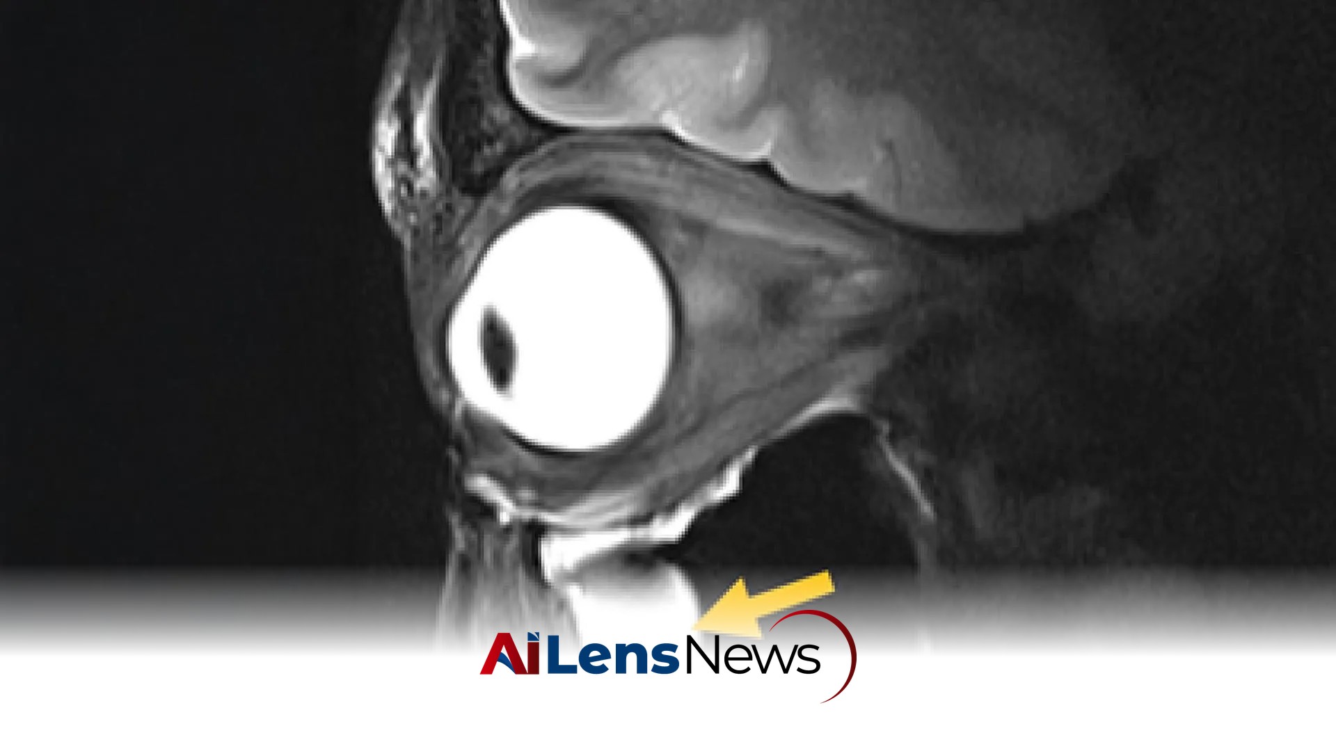


Scientists have redesigned a key piece of MRI hardware using metamaterials, allowing existing scanners to produce clearer images of difficult-to-see parts of the body in less time. This breakthrough could enhance diagnoses and improve patient comfort.
New Delhi, India Jul 10, 2026 ALN: Magnetic resonance imaging (MRI) has revolutionized the field of medical diagnostics since its inception in the late 20th century. This non-invasive imaging technique uses powerful magnetic fields and radiofrequency waves to create detailed images of the inside of the body, allowing care professionals to diagnose a variety of conditions without the need for invasive procedures. However, even with the advancements in MRI technology, certain areas of the body remain challenging to image effectively. Deep brain structures and the delicate tissues of the eye and surrounding orbit are particularly problematic due to the limitations of existing MRI hardware, which can struggle to produce clear images from these complex anatomical regions.
In a significant advancement, a team led by Nandita Saha, a doctoral student in Professor Thoralf Niendorf's Experimental Ultrahigh Field Magnetic Resonance laboratory at the Max Delbrück Center, has developed a groundbreaking MRI antenna utilizing advanced engineered materials. This innovation not only enhances the clarity of images produced but also reduces the time required for scanning, making it a potentially transformative development for the field of medical imaging. The findings from this research were published in the prestigious journal Advanced Materials.
The collaborative effort involved experts from various fields, including MRI physics, clinical ophthalmology, and translational imaging, from both the Max Delbrück Center and Rostock University Medical Center. Researchers at Rostock University are also engaged in validating this new technology for future clinical applications, which could significantly impact patient care and diagnostics.
According to Niendorf, who serves as the senior author of the paper, the integration of metamaterials into MRI antennas has led to a more efficient way of guiding radiofrequency fields. This represents a notable advancement in the application of physics to medical imaging, potentially resulting in faster and clearer MRI scans that could benefit patients across a variety of clinical contexts. The implications of this research extend beyond mere image clarity; they hint at a future where MRI technology can provide deeper insights into the physiological and pathological processes occurring within the body.
Metamaterials Improve MRI Performance
The fundamental working principle of MRI scanners involves sending radiofrequency (RF) signals into the body while simultaneously applying a powerful magnetic field. As the body's tissues respond to these signals, the scanner collects the necessary information to generate an image. Generally, stronger signals yield clearer and more detailed scans. However, traditional MRI antennas, also known as RF coils, often encounter difficulties in collecting sufficient signals from tissues that are located deep within the body or in regions with complex anatomy. Consequently, this can lead to compromised image quality and longer scanning sessions.
To address these challenges, the research team incorporated metamaterials directly into the MRI antenna design. Metamaterials are artificially structured materials engineered to interact with electromagnetic waves in unique ways that natural materials cannot. In laboratory tests, the new antenna demonstrated a remarkable ability to strengthen signals from targeted tissues, enhance spatial resolution, improve image sharpness, and accelerate data collection. These enhancements are particularly relevant for imaging delicate structures such as the eye and its surrounding tissues.
One of the significant advantages of this new antenna design is its compatibility with existing MRI equipment. This compatibility means that care facilities can implement this technology without the need for costly new infrastructure, making it a more accessible option for improving imaging capabilities. In practical applications, the researchers tested the design by imaging the eye and orbit in volunteers using a powerful 7.0 Tesla MRI scanner, a significant advancement in ophthalmological imaging.
Professor Oliver Stachs, a co-author of the paper from University Medicine Rostock, emphasized the relevance of this research for ophthalmological applications. He noted that the technology could facilitate anatomically detailed, high-spatial resolution MRI scans of the eye, potentially opening a window into the eye's physiological processes that have previously been difficult to access. Such advancements could lead to better understanding and treatment of ocular diseases.
Potential Beyond Eye Imaging
While the initial focus of the research has been on improving eye imaging, Saha highlights that the technology could have broader applications. For instance, the design could be adapted to protect sensitive areas of the body during MRI exams by minimizing unwanted heating around medical implants, which is a concern in MRI procedures. Additionally, the antenna could enhance MRI-guided cancer treatments by allowing for more precise delivery of RF energy in procedures such as tumor hyperthermia or thermal tissue ablation, where targeted heating is used to destroy cancer cells.
Faster Scans and Better Diagnoses
The duration of MRI exams can often be lengthy and uncomfortable for patients, especially when repeated scans are necessary due to inadequate imaging of critical anatomical details. The new antenna's ability to produce clearer images more quickly has the potential to shorten scan times, thereby improving patient comfort and increasing the confidence of physicians in their diagnoses. Furthermore, the compact and lightweight design of the antenna allows for customization to suit different body parts, which could further enhance patient comfort during imaging procedures.
Niendorf expressed optimism about the future adaptability of this design, suggesting that it could eventually be tailored for MRI systems operating at both lower and higher magnetic field strengths than the 7.0 T used in their tests. Beyond the eye and brain, the technology could also be applied to imaging other organs, such as the heart and kidneys, or utilized in monitoring metabolic processes and tracking the distribution of pharmaceuticals within the body.
Moreover, the technology holds promise for improving specialized MRI techniques that focus on imaging elements other than hydrogen, such as sodium and fluorine. By generating stronger signals and producing higher quality images, these advancements could enhance the diagnostic capabilities of MRI in a variety of clinical scenarios.
Dr. Ebba Beller, another co-author of the paper from Rostock University Medical Center, remarked on the transformative potential of innovations in imaging hardware, asserting that this study represents an important step toward the development of next-generation MRI technology.
Next Steps
Looking ahead, the research team plans to conduct larger clinical studies involving multiple hospitals to further validate the effectiveness of the new antenna design. They are also working on modifications to extend its application to additional organs, including the heart and kidneys, which could broaden the scope of its clinical utility. The ongoing collaboration between Stachs and Niendorf is set to continue, with reciprocal visiting scientist appointments fostering further research and development in this promising area of medical imaging.
The project received funding from the German Research Foundation (DFG) as part of a joint collaboration between the Max Delbrück Center and the Medical University Rostock, highlighting the importance of interdisciplinary approaches in advancing medical technology.
In conclusion, the development of this new MRI antenna represents a significant advancement in medical imaging technology. By leveraging the principles of metamaterials, the research not only enhances the quality and speed of MRI scans but also opens up new avenues for clinical applications that could ultimately lead to better patient outcomes and more effective diagnostic practices.
To learn more about the latest developments in Medical Innovations, stay updated with our exclusive reports and analyses on AiLensNews.